


I would like to welcome you to the first fully digital batch of papers from the Society of Radiographers’ Insight journal. This newest release is packed with insightful submissions all centred on how contemporary radiography is shaped through clinical and academic innovations.
Leigh-Anne Keyte and Marie Cook provide an audit assessing the value of bone loss on projection radiography as a risk factor for DXA scan referrals. The authors suggest that projection radiography reports need to be combined with clinical information on referral forms and reported risk factors on patient questionnaires as the best approach to determining appropriate referrals for DXA scans. This would ensure the right patients for DXA are identified, minimise variation in the referral process, optimise utilisation of scanners, and reduce unnecessary radiation on ineligible patients.
Two separate case reports are also presented highlighting how advancing roles in radiography contribute to proper identification, diagnosis and ongoing management for infrequent pathologies. Helen Watts reports on sub-acute/chronic paediatric osteomyelitis, a condition of limited occurrence in children. She highlights the importance of prompt imaging referral, acquisition and reporting, as well as result escalation for optimum patient management. Following on, the case by Rahul Modi centres on neuroimaging features of non-ketotic hyperglycaemia, with correlation of CT and MRI findings. The authors emphasise the need for radiographers’ awareness of metabolic stroke mimics, and how CT and MRI investigations can support appropriate patient triage, diagnosis, and management.
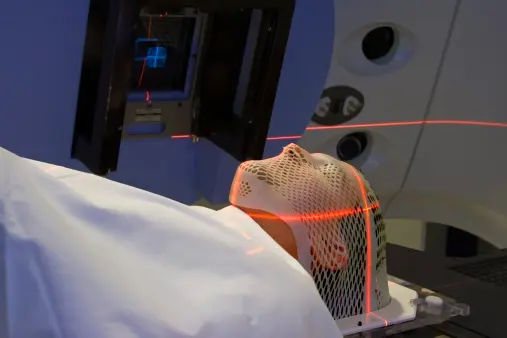
I am personally drawn to the paper by Liam Bunyan, an original pre- and post-survey research assessing how virtual radiotherapy simulation can impact students' understanding. The authors found an increase in self-reported knowledge of key concepts across all measured domains. Such a low-cost education intervention, capable of being adopted outside the clinical setting, can provide versatility in the training of radiography students, especially in low-resource settings. Although virtual simulation teaching should not replace conventional education approaches, there is a need to establish longitudinally how a combination of both performs when compared to either for student training. This should be a key area for future research.
The readers are also treated to reflective reviews focusing on two separate career trajectories for newly qualified radiographers. Alexandra Partner provides an experiential account of the NIHR INSIGHT programme. With the radiography profession already on the back foot in research engagement compared with most medical and health professions, the authors’ detailed insight on this programme could facilitate uptake and boost clinical academic careers for radiographers. Also, Hannah Chandler draws on their experiences as both a nuclear medicine practitioner and educator to provide valuable insights about the profession. The author highlighted reasons why radiography students are not attracted to the modality after graduation: limited exposure, misconceptions and lack of proper mentorship.
The narrative review by Kalpana Lakhani argued that variability in polycystic ovarian syndrome diagnosis using ultrasound is enabled by both technical limitations and governance failures. They advocate for consistent and standardised techniques among ultrasound practitioners to minimise delayed definitive diagnosis of the syndrome and unnecessary repeat imaging. Finally, a pictorial review by Shagufta Fazal explores how in-vivo use of Hyperpolarised Xenon (129Xe) lung MRI technique can critically address most of the limiting factors affecting conventional lung MRI investigations and diagnosis.
I am confident that readers will find valuable learning points in all papers on this platform, specifically on the need to develop beyond technical image acquisition towards evidence-informed decision-making, continuous professional learning, research engagement and adaptation to emerging innovations.
About the authors
Edozie Iweka is an advanced practice radiographer for clinical trials and research, and a clinical specialist in CT and MRI. He is currently a Wellcome Trust GW4CAT HP doctoral fellow with the University of Bath, and his research interest spans across cross sectional imaging in oncology care, AI innovation in medical imaging and evidence synthesis.
.jpg?format=webp)